Location: MRBIII U3202

ImageQuant LAS 4000 delivers:
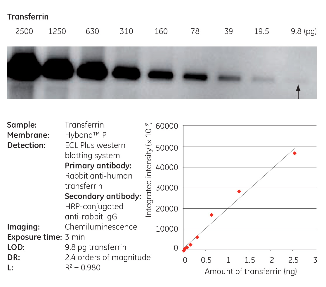
- Wide linear dynamic range: Capture true 16-bit images and precisely quantitate weak and strong signals over three orders of magnitude in the same image, avoiding time-consuming and costly multiple film exposures. This is in stark contrast to film which has a dynamic range that is non-linear and is 1.5 orders of magnitude or less.
- High resolution: Accurate quantitation of blots up to 21 × 14 cm in size. The 3.2 MPixel Fuji CCD chip (11 um pixel size) can deliver up to 6.3 Mpixel image resolution, achieved via a unique diagonal pixel configuration (see image of chip design below).
- Low noise: The camera is typically cooled to -25°C (maximum -35°C) by a two-stage thermoelectric module, enabling longer exposure times. This provides users with less background, which is especially important for precise quantitation of very weak signals in chemiluminescent Western blotting.
- High sensitivity: ImageQuant LAS 4000’s camera is fitted with a large aperture FUJINON F0.85 43 mm lens, one of the best in the industry. This allows the instrument to capture chemiluminescent signals on a Western blot down to picogram levels of target protein. Sensitivity may be increased by binning of up to 8 x 16 pixels. The camera has automatic or manual capture modes and exposure times (1/100 s to 30 hours).
- Uniform quantitation: Using a scanner to scan a film for quantification can introduce artifacts and further complicate the quality of your results. The LAS 4000 has distortion, dark frame, and flat frame corrections which are applied to each imaging mode for optimal precision.
- ImageQuant TL: GE’s quantitation software allows you to click one button to find your lanes and bands. A couple more steps and you’ve subtracted your background and quantitated your bands in the time it took you to draw boxes around your bands in ImageJ!
IQ LAS 4000_verAB DataFile.pdf
GE Healthcare’s unique CCD chip design with octagonally-arranged pixels.
![]()
Quantification down to picogram concentrations
GFP Imaging
In order for GFP to fluoresce, it needs to remain in a semi, non-denaturing condition. There are a number of ways to do this depending on the actual application:
- Non-denaturing gel
- *SDS-PAGE gel without boiling the GFP sample
- #Suspension of the cells in solution in a 96-well plate
- Directly imaging bacterial colonies expressing the GFP-fusion proteins in a Petri dish.
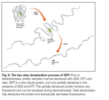
*Partial denaturation of GFP by SDS will still keep the fluorescence of the protein fairly strong. What needs to be avoided is boiling the sample. Heating will denature the GFP protein to a point that will no longer fluoresce (see Figure 6 below).
#Always ensure you use plastic or glass that has the “LF” designation (low-fluorescence). As long as you use the proper plastics or glass you should not have any problems whatsoever with any unwanted fluorescence or glare.