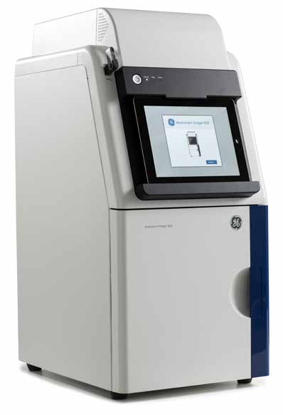
AI600 Chemiluminescent Imager
Location: U3202 MRB3
Reservations Optional on VU iLab website
Billing is determined by number of scans written on the log sheet located near the instrument.
The Log sheet must be filled out legibly and in full.
A reservation is not required for this instrument. However, if you do not have a reservation and someone comes to use the instrument and they do have a reservation, then you must remove your sample and give them immediate access.
Amersham Imager 600
 Amersham Imager 600 series is a new range of sensitive and robust imagers for the capture and analysis of high resolution digital images of protein and DNA samples in gels and membranes. These multipurpose imagers bring high performance imaging to chemiluminescence, fluorescence, and colorimetric applications. The design of Amersham Imager 600 combines our Western Blotting application expertise with optimized CCD technology and exceptional optics from Fujifilm™. The system has an integrated analysis software and intuitive workflow, which you can operate from an iPad™ or alternative touch screen device, to generate and analyze data quickly and easily.
Amersham Imager 600 delivers:
• Intuitive operation:
You can operate the instrument from a tablet computer with an intuitive design and easy-to-use
image analysis software. You do not need prior imager experience or training to obtain high-quality results. Use the automatic capture mode for convenient exposure
• Excellent performance:
The system uses a superhoneycomb CCD and a large aperture f/0.85 FUJINON™ lens, which consistently delivers high-resolution images, high sensitivity, broad dynamic range (DR), and minimal cross-talk
• Robustness:
Combining minimal maintenance with our proven expertise in Western blotting and electrophoresis makes the imager well suited for multiuser laboratories. Amersham Imager 600 is an upgradable series of imagers that can grow with your imaging needs
Description
Amersham Imager 600 series is equipped with a dark sample cabinet, a camera system, filter wheel, light sources, and a built-in computer with control and analysis software. Network connection and USB ports are standard (Fig 2). Settings such as focus, filter, illuminator, and exposure type are automatically controlled by the integrated software. You would obtain high resolution images and precise quantitation of low signals with the multipurpose 16-bit 3.2 megapixel camera fitted with a large aperture lens. The detector is cooled to reduce noise levels for high sensitivity and wide dynamic range. Rapid cooling leads to a short startup time, which makes the instrument ready to use in less than 5 min. You can place the sample tray at one of two different heights
in the sample compartment to produce image-acquisition areas of 220 × 160 mm and 110 × 80 mm, respectively.