>
Vanderbilt University
Vanderbilt University
Menu
The Lee Lab at Vanderbilt
Home
Our Research
People
Current Lab Members
Past Lab Members
Publications
Lee Lab News
Lab Fun
Contact Us
Links
Positions
Close Menu
Vanderbilt University
The Lee Lab at Vanderbilt
All Vanderbilt
Home
Our Research
People
Current Lab Members
Past Lab Members
Publications
Lee Lab News
Lab Fun
Contact Us
Links
Positions
About
About Vanderbilt
History
Quick Facts
University Leadership
Contact
Academics
Academics Overview
Program Finder
Admissions
Undergraduate Admissions
Graduate & Professional School Admissions
Cost & Financial Aid
Athletics
Campus Life
Life at Vanderbilt
Nashville
Our Campuses
Housing & Dining
Organizations & Community
Health & Safety
Research
News
Uncategorized
Mouse embryonic fibroblast (MEF) cells stained for beta-catenin (red), actin (yellow), and DNA (blue).
June 12, 2018
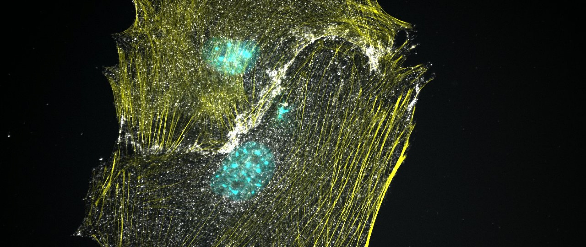
Casein Kinase 1alpha null MEF cells exhibit constitutively active Wnt signaling. Cytosolic beta-catenin (white) localization has now been shifted to the nucleus (blue). Beta-catenin also remains localized at cell-cell contact as part of the cadherin complex. Actin is labeled in yellow.
June 12, 2018
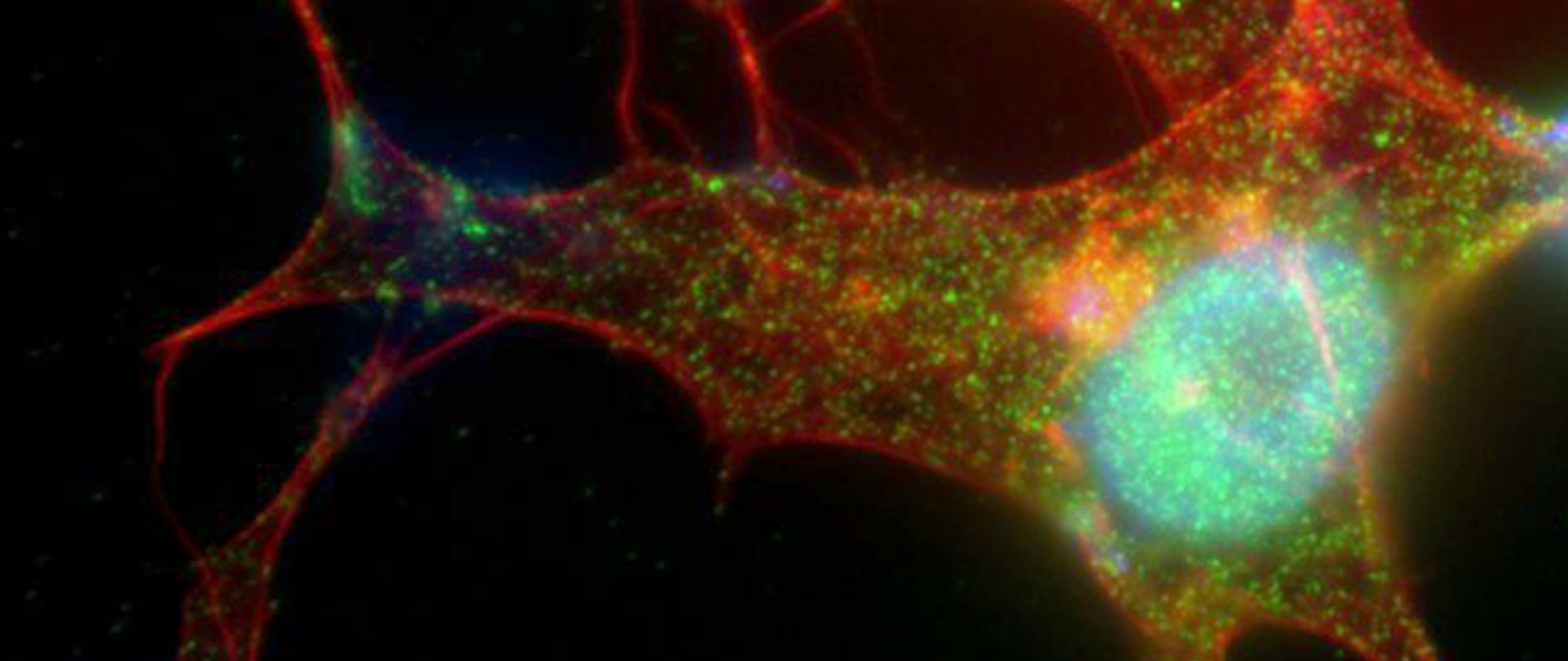
Retinal pigmented epithelial (RPE) cells are highly responsive to Wnt ligand stimulation. Intense beta catenin (green) localization in the nucleus (blue) is observed upon treatment with Wnt3A. Actin is labeled in red.
June 12, 2018
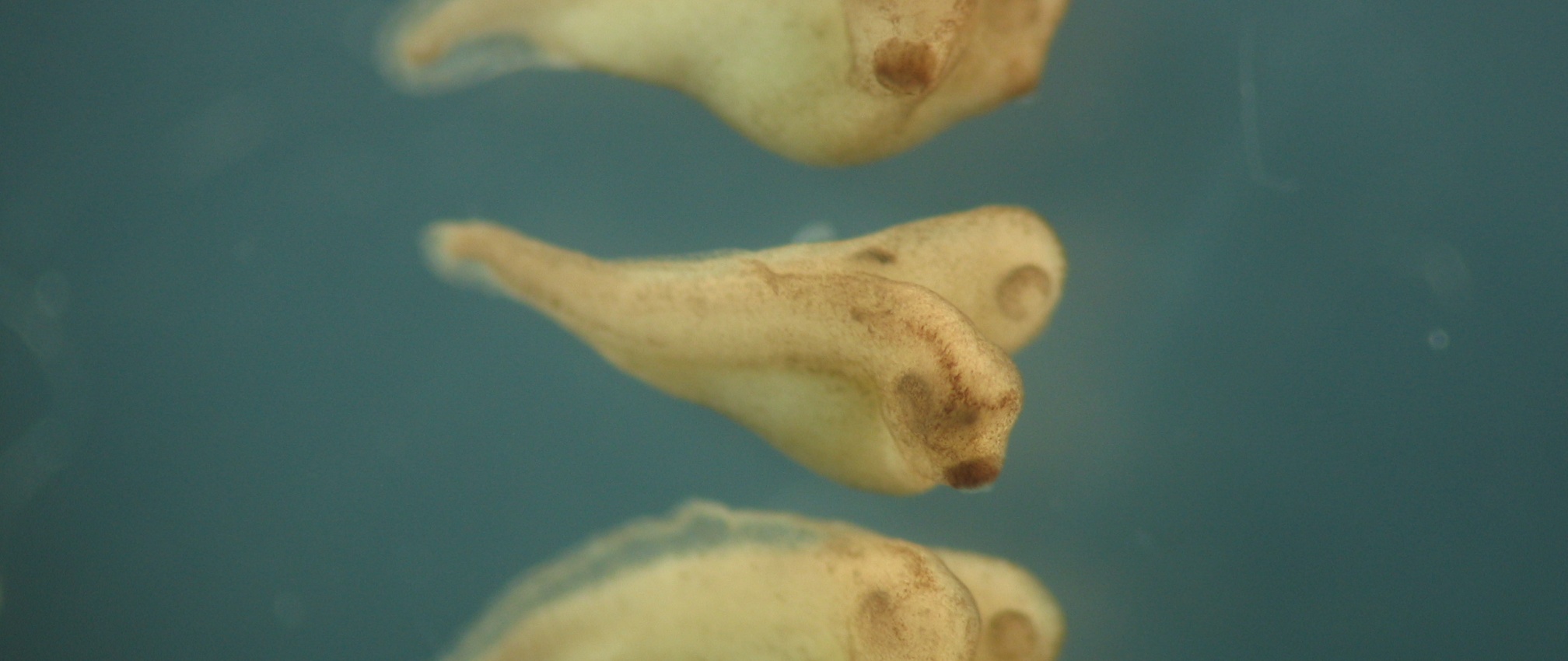
Xenopus laevis injected with beta-catenin, a critical component of the Wnt pathway, at the 4-cell stage results in embryos with duplicated axes.
June 12, 2018
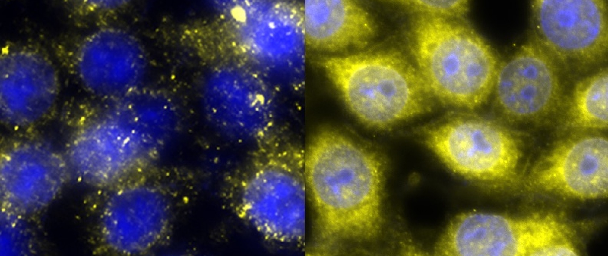
APC is a tumor suppressor that negatively regulates the Wnt pathway. RKO cells with wild-type (left) and knocked out APC (right). These cells have been stained for beta-catenin (yellow) and DNA (blue) Note the intense staining of beta-catenin upon loss of APC.
June 12, 2018
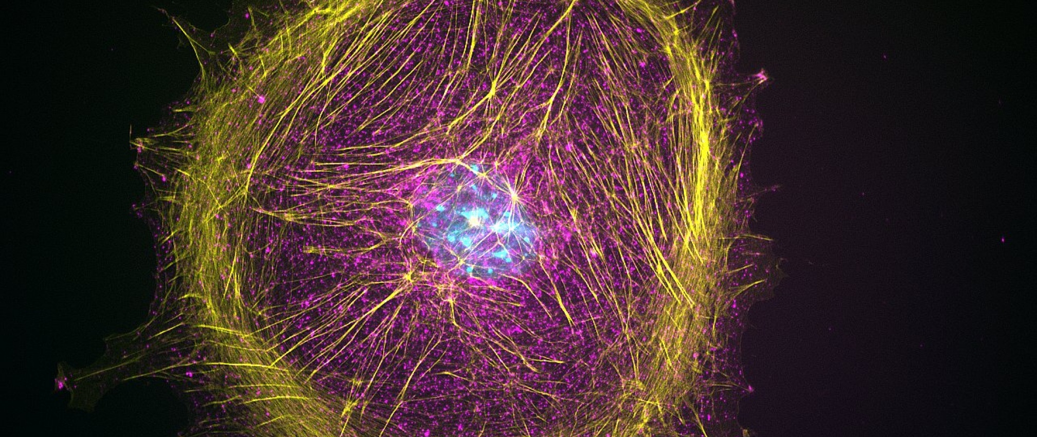
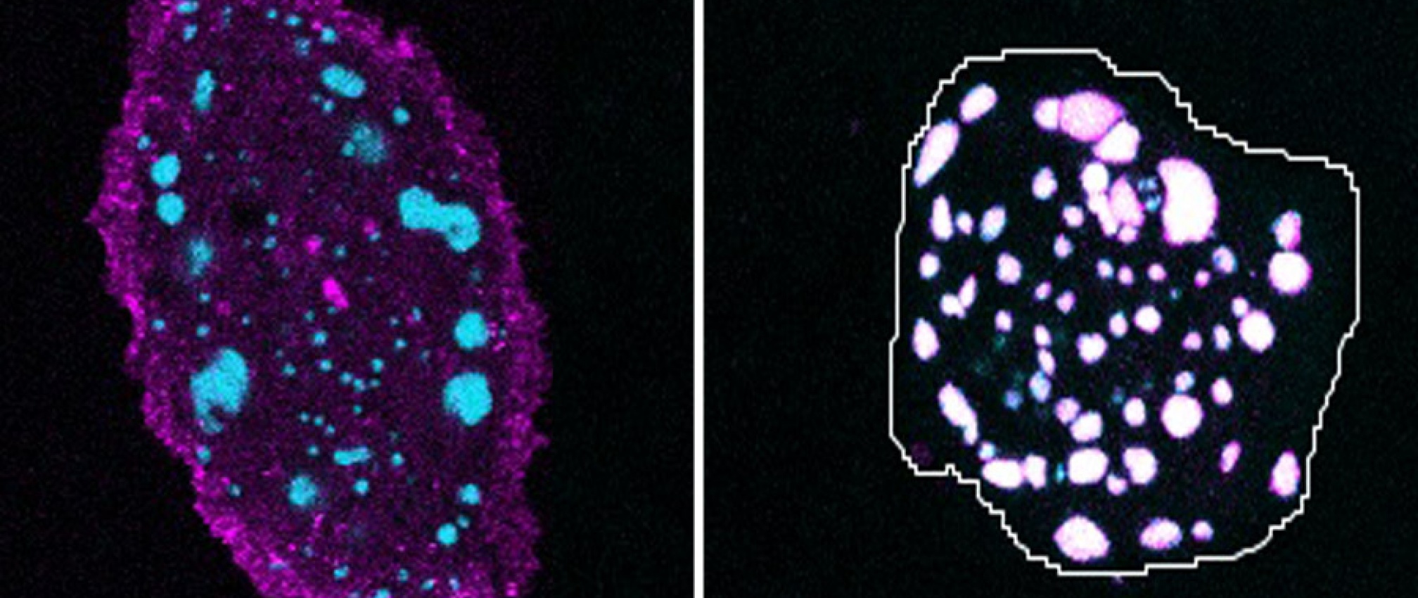
LRP6 (magenta) is internalized and colocalizes with Axin (cyan) upon Wnt ligand activation. Left panel shows untreated RPE cells. Right panel shows RPE cells treated with Wnt3a.
June 12, 2018
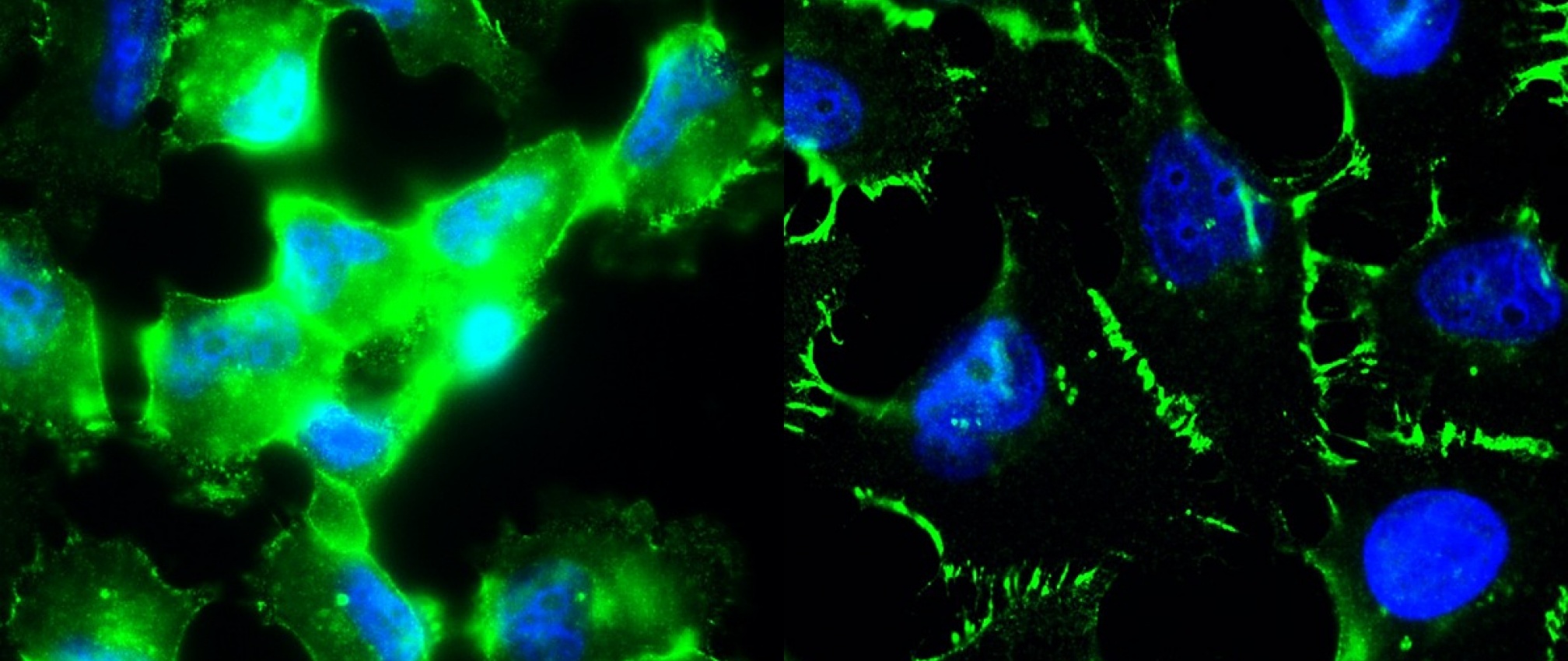
The triple negative breast cancer line, MDA-MD-231, exhibit high levels of beta-catenin staining (left) and elevated Wnt signaling. Incubating MDA-MD-231 cells with an antibody that binds the Wnt coreceptor, LRP6, dramatically decreases cytoplasmic beta-catenin staining and promotes beta-catenin localization to the cell junctions (right). Green is beta-catenin and blue is DNA.
June 12, 2018
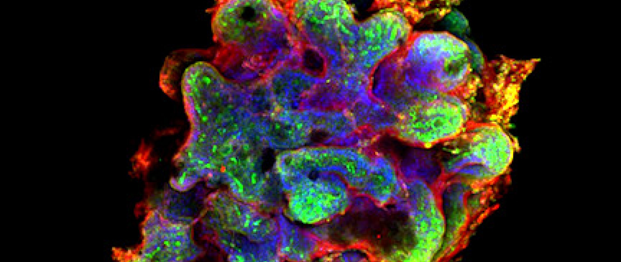
Human intestinal organoids generated from IPS cells. Beta-catenin is (red), actin (green) and DNA (blue).
June 12, 2018
Intestinal enteroids represent a valuable ex vivo system that are reminiscent of normal gut epithelium. These enteroids contain stem cells and produce cell types found within the intestinal epithelium. Enteroids are being used to study the GI tract in normal and pathological situations as well as screening for drugs.
June 12, 2018
Previous
Next