Research
In the Bowden Lab, we take an interdisciplinary approach to research that combines knowledge and ideas from optics, electrical engineering, computer vision, machine learning, microfluidics, nanophotonics and other fields to develop new technologies with the potential for clinical translation. Many of our works focus on technology development for a specific imaging technology: optical coherence tomography; a second research thrust is aimed at technology development for resource-constrained environments, such as for applications to global health, rural health, and personal health.
Optical Coherence Tomography (OCT)
OCT is an optical imaging technique that provides high-resolution imaging of subsurface tissue structures. One way to think about it is as an optical analog to ultrasound. OCT is one of the core technologies used in the BBOL. Our work in this area includes development novel software and hardware and applying OCT to various clinical applications.
Novel system development
Our lab has introduced several new system designs that advance the state-of-the art of OCT. These enhancements include aspects such as improved system speed, improved imaging depth and increased flexibility.
- Combined optical coherence tomography and hyper-spectral imaging using a double-clad fiber coupler
- Wide-field, full-field optical coherence microscopy for high-axial-resolution phase and amplitude imaging
- Polarization-sensitive interleaved optical coherence tomography
- Single-shot speckle noise reduction by interleaved optical coherence tomography
Novel algorithms
Extracting meaningful, quantitative information from image data often requires new and advanced algorithms that exploit knowledge of the underlying system architecture to interpret what the data mean. We have developed several algorithms that push the boundaries of what we can learn from OCT data.
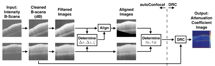
- Automatically determining the confocal parameters from OCT B-scans for quantification of the attenuation coefficients
- Classification of basal cell carcinoma in human skin using machine learning and quantitative features captured by polarization-sensitive optical coherence tomography
- Enhanced depolarization contrast in polarization-sensitive optical coherence tomography
- Iterative re-weighted approach to high-resolution optical coherence tomography with narrow-band sources
- Automated, Depth-resolved Estimation of the Attenuation Coefficient From Optical Coherence Tomography Data
- Automated mosaicing of feature-poor optical coherence tomography volumes with a freehand dual-modality probe
Calibration and tissue-mimicking phantoms

Development of new technologies can sometimes be hampered by access to appropriate reference and measurement standards. Our lab has pioneered a number of new techniques to produce phantoms that facilitate OCT system development.
- Multimodal 3D cancer-mimicking optical phantom
- Fabrication of Healthy and Disease-mimicking Retinal Phantoms with Tapered Foveal Pits for Optical Coherence Tomography
- Three-dimensional, distendable bladder phantom for optical coherence tomography and white light cystoscopy
- Variable-sized bar targets for characterizing three-dimensional resolution in OCT
Clinical Applications

As the ultimate goal of our is clinical translation, we work closely with clinicians to apply our technologies to unmet needs such as early cancer detection in the bladder and skin, cancer treatment, infertility, and other areas.
- Registration of free-hand OCT daughter endoscopy to 3D organ reconstruction
- Classification of basal cell carcinoma in human skin using machine learning and quantitative features captured by polarization-sensitive optical coherence tomography
- Two-dimensional cochlear micromechanics measured in vivo demonstrate radial tuning within the mouse organ of Corti
- Label-free characterization of vitrification-induced morphology changes in single-cell embryos with full-field optical coherence tomography
- Rapid scanning catherscope for expanded forward-view volumetric imaging with optical coherence tomography
- Noninvasive in vivo imaging reveals differences between tectorial membrane and basilar membrane traveling waves in the mouse cochlea
- Automated identification of basal cell carcinoma by polarization-sensitive optical coherence tomography
Technology for resource-constrained environments
Resource-constrained environments provide fodder for creative ideas.
Point-of-care urinalysis

Urine is one of the most easily accessible body fluids and contains an enormous amount of information about the state of health. We have implemented a number of technologies aimed at making measurements of urine contents robust and easy-to-perform in the hands of a non-specialist.
- Low-power, low-cost urinalysis system with integrated dipstick evaluation and microscopic analysis
- Robust dipstick urinalysis using a low-cost, micro-volume slipping manifold and mobile phone platform
- Label-free and noncontact optical biosensing of glucose with quantum dots
- Quantifying colorimetric assays in paper-based microfluidic devices by measuring the transmission of light through paper
Magnetic Levitation
Diamagnetic objects suspended in a paramagnetic medium can be levitated by balancing magnetic forces against gravitational forces; the physical position of objects in this context is determined by the density of the object. MagLev systems are cheap to implement and extremely sensitive.
- Using Magnetic Levitation for Non-Destructive Quality Control of Plastic Parts
- Non-contact Orientation of Objects in Three Dimensional Space Using Magnetic Levitation
- Templated Three-Dimensional Self-Assembly Using Magnetic Levitation
- Using Magnetic Levitation for Three Dimensional Self-Assembly