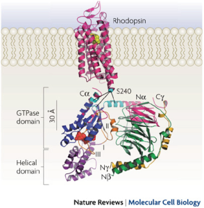
 Ribbon model of rhodopsin (Protein Data Bank (PDB) ID 1GZM12) juxtaposed with the Gt heterotrimer (PDB ID 1GOT32). G-protein-coupled receptors (such as rhodopsin; pink) share a common structure with seven-transmembrane-spanning α-helices. Receptor activation exposes the G protein binding site that is formed by the intracellular loops of the receptor. Biochemical studies have identified several receptor contact sites on the G protein (pink and cyan). A few studies have also defined specific point-to-point interactions, such as between Ser240 on intracellular loop 3 of the receptor (cyan sphere) with specific regions of the Gα N terminus (Na), C terminus (Ca) and α4–β6 loop (cyan)66, 70. These contact regions, with the fatty acid modifications on the Gα N terminus and Gγ C terminus, suggest a probable orientation with respect to the membrane. This orientation places GDP (red spheres) in the nucleotide-binding pocket on Gα (blue) ~30 Å from the sites of nearest receptor contact, posing the question of how receptors cause GDP release from this distance.