Inter-relationships & Functions of MT-binding Proteins » Rassf1A homepage_b
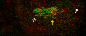
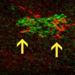
Image showing two distinct localizations of the MT-binding protein RASSF1A: segmental RASSF1A localization to peripheral MTs (indicated by white arrow), and RASSF1A-bound Golgi-derived MTs (indicated by yellow arrows). The Golgi is shown in green, RFP-RASSF1A is shown in red.


Leave a Response
You must be logged in to post a comment