Perfusion Imaging
Ultrasound is excellent at visualizing flow in large arteries and veins, but as the vasculature gets smaller and the blood velocity decreases ultrasound can no longer detect the flow as in our example below. We’re interested in visualizating slow blood flow, but more specifically, we’re interested in using ultrasound to visualize perfusion in unresolvable vasculature without contrast agents. For a long time this was considered to be impossible, but thanks to some recent work by our group and others, we’re showing that it can be done.
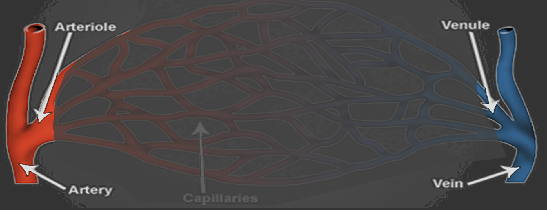
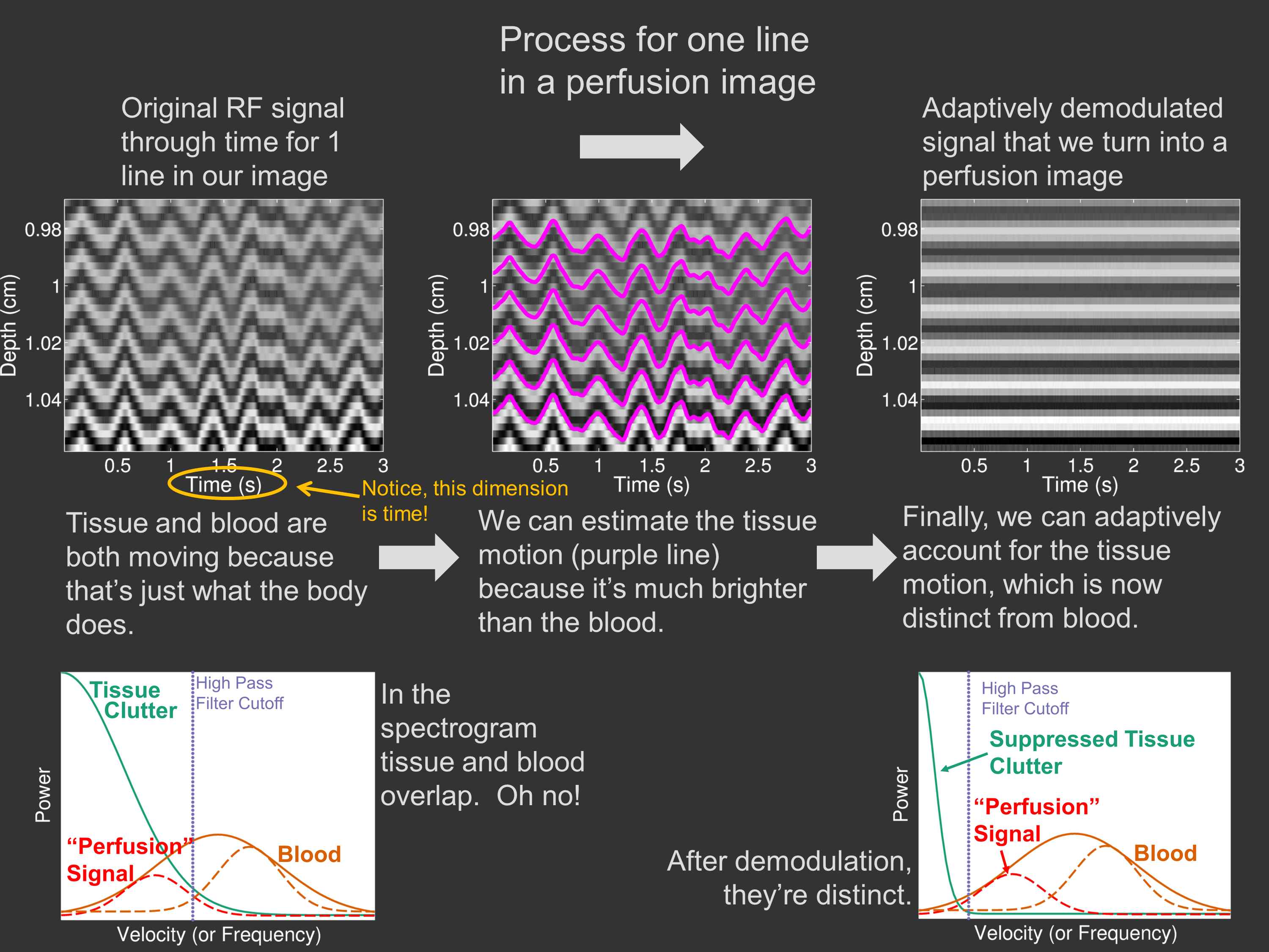
The challenge is that the motion of the slow moving blood that we’d like to characterize is moving at about the same speed as the surrounding tissue. This means that the ultrasound signal from tissue and blood are very similar and can’t be differentiated using normal methods. We’ve shown that by directly measuring the motion of the tissue–something that ultrasound can do very well–we can better separate the tissue and the blood.
We’ve used this to visualize small changes in blood flow from muscle contraction, and more recently we’ve shown that this can be used to help physicians with certain procedures–more to come soon!
The example below is from one of our muscle contraction studies.