NeuroImaging Outside the Brain Lab (NOB-L)
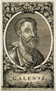 Galen of Pergamum (130-200AD), the attending physician to Marcus Aurelius, recounted an anecdote of how a sophist Pausanias was injured falling off his horse. Soon after his injury, Pausanias lost sensation in his fingers, but did not lose motor function. At the time, Pausanias was treated with various medications to treat his hand, but none worked. Galen decided that it would be better to treat the site of injury (in this case, C7) with the same medications, and recounted that his treatment of Pausanias resulted in a complete recovery. Galen attributed this to the impact of the spinal cord on the motor and sensory function throughout the body and that the spinal cord is integrally involved in the function of the human being. He stated that it is better to treat the site of pathology rather than its “somatic manifestations”. Thus, 2000 years ago, it was understood that the small nerves throughout the body, and injury of such nerves, were integral to day-to-day function.
 Following this, a study performed nearly 200 years ago (Caswell, Illustrations of the Elementary Forms of Disease, 1838) discovered that the spinal cord bears a significant burden of multiple sclerosis lesions that may drive neurological deficits in the disease. In fact, several studies have shown that the relationship between spinal cord damage and neurological performance may be stronger than considering lesions in the brain alone.
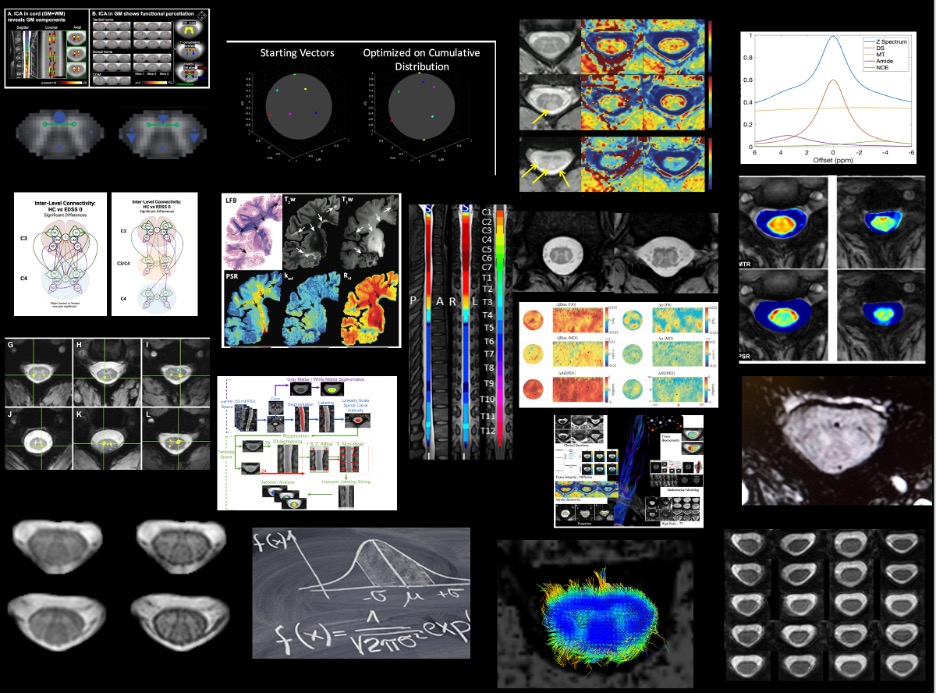
It is now well understood that developing tools that could assess the health and welfare of small nerves of the human body may have a significant impact in understanding the health and welfare of the human being in many different nervous system diseases, syndromes, and injury. However, non-invasive radiological measures such as MRI are often not robust, reliable, well-developed, informative, or other non-useful terms. MRI measures of these small nerves struggle to achieve the same diagnostic and prognostic impact that similar radiological measures do for other tissues in the body.
 In 1946, the great Philosopher Irving Berlin said that “Anything you can do (I can do better)” and serves as one of the motivations for the NOB-L lab. There is a tremendous wealth of impressive literature that shows how novel, quantitative, high-resolution, high-fidelity, high-specificity MRI markers can be performed in the brain providing the scientific and clinical community a rich field to harvest for understanding diseases and disease manifestations. However, often similar tools for the study of the spinal cord, the optic nerve, peripheral nerves, etc. are either in their infancy, or are not well suited for providing the same information that can be obtained in the brain. In our lab, we seek to address that challenge by developing similarly useful techniques that have been explored in the brain for the small nerves of the human body.
Thus, our lab seeks to do 3 things:
- Develop, optimize, and evaluate advanced neuroimaging techniques and technologies for the small, somewhat understudied tissues of the human nervous system.
- Apply the developed tools to the study of diseases, syndromes, and injury of the human nervous system to understand the pathological manifestations of such conditions.
- Improve imaging for the direct purpose of improving a clinician’s ability to diagnose, treat, evaluate, and prognosticate so that health care for the human condition can be strengthened. We aim for our techniques and technologies to be useful, usable, and used, clinically.