Welcome
The spatial analysis of complex proteomes, lipids and metabolites in tissues samples provides information that can facilitate the detection of disease states, responses to therapy and drug toxicity.
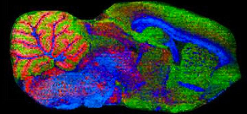
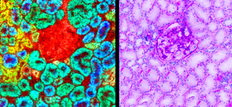
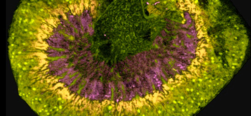
Imaging mass spectrometry (IMS), pioneered at Vanderbilt, is done by analyzing mass spectral data acquired across a grid within a tissue section. Recent work suggests that IMS can identify biomarkers of diseases, including different cancers.
The Imaging Mass Spectrometry Core laboratory provides direct molecular profiling and imaging of intact tissues by MALDI-MS.
Spatial patterns of molecular expression can be analyzed with bioinformatics and biostatistical methods to identify sets of diagnostic spectral markers. Identification of the proteins and protein fragments that account for biomarker signals is done in collaboration with the Proteomics Core Laboratory and identification of lipids and metabolites is done in collaboration with the Mass Spectrometry Core Laboratory.
If you would like to inquire about using the Imaging Mass Spectrometry Core, please click here to request a consultation.